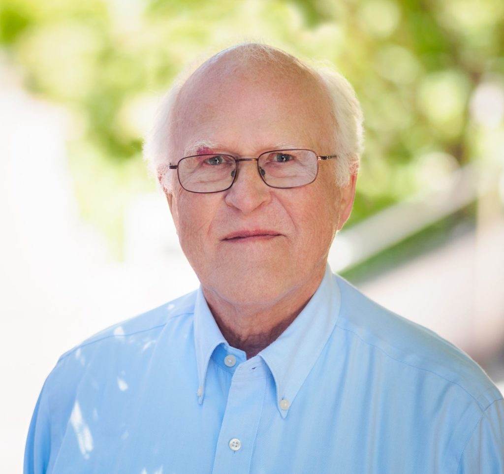
 Robert (Bob) Martin Glaeser is a Biophysicist Senior Scientist and an Emeritus Professor at UC Berkeley. During his 56-year career at the Lab, Glaeser’s work to develop the ability to successfully create high-resolution images of biological specimens has been integral to the advancement of cryogenic electron microscopy (cryo-EM). This imaging technique launched the fields of structural biology and biochemistry into an exciting new era of discovery.
In the 1960s, scientists were beginning to use electrons to image samples that had been frozen mid-motion, expanding a technique that had previously been used for dead or inanimate matter to proteins and other biomolecules. But these specimens were being damaged by radiation, preventing researchers from being able to map the structure of proteins and other key biological molecules. Radiation was not the only challenge; scientists also had to contend with marginal image quality and unsatisfactory attempts at phase contrast. However, thanks to Glaeser’s foundational work in the field, and his prolific portfolio of inventions, these problems – and many others – were solved. In fact, his solutions ensured that cryo-EM could help propel the emerging field of structural biology.
Starting in the early 1970s, Glaeser was the first to demonstrate that cryogenic cooling offered enormous gains in image quality. He quantified radiation damage in crystalline catalase and small organic molecules, noting that electron doses could be lowered by averaging over ensembles of particles. This discovery led him to demonstrate electron diffraction from frozen catalase crystals to higher than 3-Å resolution. With these key breakthroughs, Glaeser laid the foundation for what has become known as cryo-EM, and is now the preferred method for investigating the structure of biological macromolecules. His research and technologies helped an entire community to understand the physical limits of image formation. As Wolfgang Baumeister of the Max Planck Institute for Biochemistry in Germany observed, “There is hardly anybody else in the field of biological electron microscopy with a more profound understanding of the underlying physics.” Over time, Glaeser and his colleagues developed radically improved methods – from sample preparation to image recording – that paved the way to the “resolution revolution” of the 2010s and enabled scientists to intimately understand what was once thought impossible: the structure and function of macromolecules.
Internationally recognized for his critical role in cryo-EM’s formative development, Bob was cited five times by the Nobel committee in their scientific description of the 2017 Nobel Prize in Chemistry for cryo-EM. One of the three laureates, Richard Henderson of the UK’s Medical Research Council Laboratory of Molecular Biology noted that, “Bob could easily have been one of the recipients, had the committee decided to emphasize slightly different aspects of the work.” His many awards and honors include Microscopy Society of America’s 2004 Distinguished Scientist Award for the Biological Sciences, the UCLA Department of Chemistry and Biochemistry’s 2018 Glenn T. Seaborg Medal, and induction into both the National Academy of Sciences and the American Academy of Arts and Sciences.
In the last two decades, Glaeser has become intensely interested in two applied problems at either end of the cryo-EM process: the unreliability of sample preparation and the failure to achieve the maximum theoretical phase contrast in the imaging process. His focus on these issues attracted the attention of the world’s leading cryo-EM companies: FEI Company, Gatan, and ThermoFisher Scientific, where they use this method to provide key information on drug targets and disease mechanisms. His partnerships with these companies has led to five patent applications in the last five years, and he has filed 18 invention disclosures over the last 25 years.
Thanks to Glaeser and other colleagues who have collaborated on continuous improvements to this methodology, Berkeley Lab is unique across the DOE national laboratory complex in its cryo-EM capabilities. And this year, he helped attract an $11 million grant from the Chan Zuckerberg Initiative for laser-based phasecontrast for next-gen proteomics in cryo-EM.
In addition to his focus on the science and technology of imaging, Glaeser has worked to recruit and build a community of structural biologists focused on electron microscopy and cryo-EM. He has also mentored, trained, and taught many students and postdoctoral scientists. These efforts have ensured that Berkeley Lab has been among the best equipped and best staffed institutions working on the three dimensional electron microscopy of biological structures. As his colleague Eva Novales put it, Glaeser’s contributions “pointed the field in the right direction to arrive at the present time, when cryo-EM is a blooming technique and considered by most [to be] among the most exciting and revolutionizing methodologies in molecular biology.”
